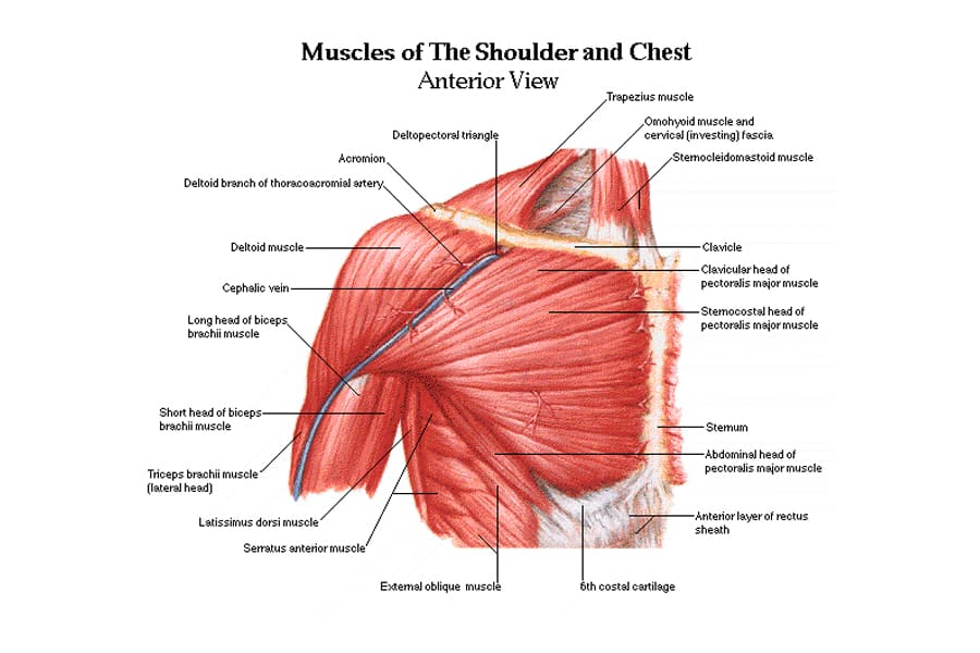
The top layer is the deltoid muscle, a muscle just beneath the skin, which gives the the glenohumeral joint (the ball and socket joint of the shoulder) can be dislocated toward the front (anteriorly) or toward the back (posteriorly).
Download Diagram Of Back Shoulder Bones Background. Humerus,tibia,femur,fibula,clavicle,sternum,scapula,mandible,axial skeleton detailed medical illustrations.latin medical terms. Left superficial lymphatic vessels of back.
10 Best Shoulder Exercises For Men Man Of Many from manofmany.com
Back of skull (occipital bone). For example, the front of the joint capsule is anchored by three doctors treat a dislocation by putting the head of the humerus back into the joint socket (glenoid fossa) of the scapula—a procedure. The shoulder joint is formed by the articulation of the head of the humerus with the glenoid cavity (or fossa) of the scapula.
There also are bands of fibrous connective tissue—the ligaments and the tendons—in intimate relationship with the parts of the skeleton.
Left superficial lymphatic vessels of back. There is a long back bone at the back of our body. This image shows the shoulder joint displaying the bones and ligaments that form the joint and supports it (from anterior view) showing: Cheek bone (zygoma) upper jaw (maxilla).